

Description
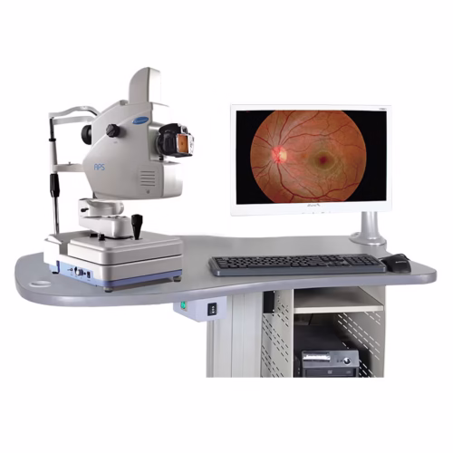
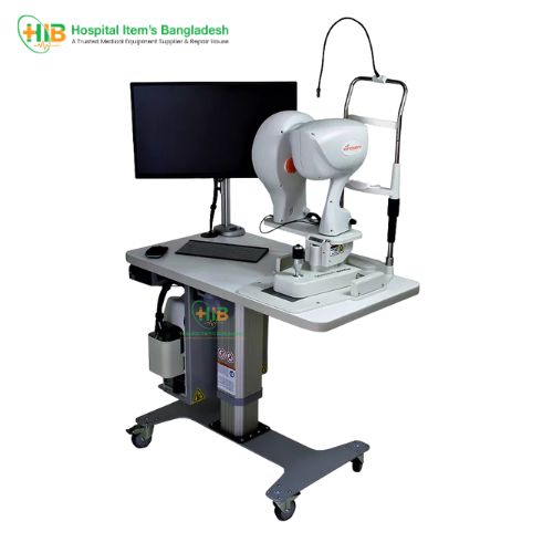
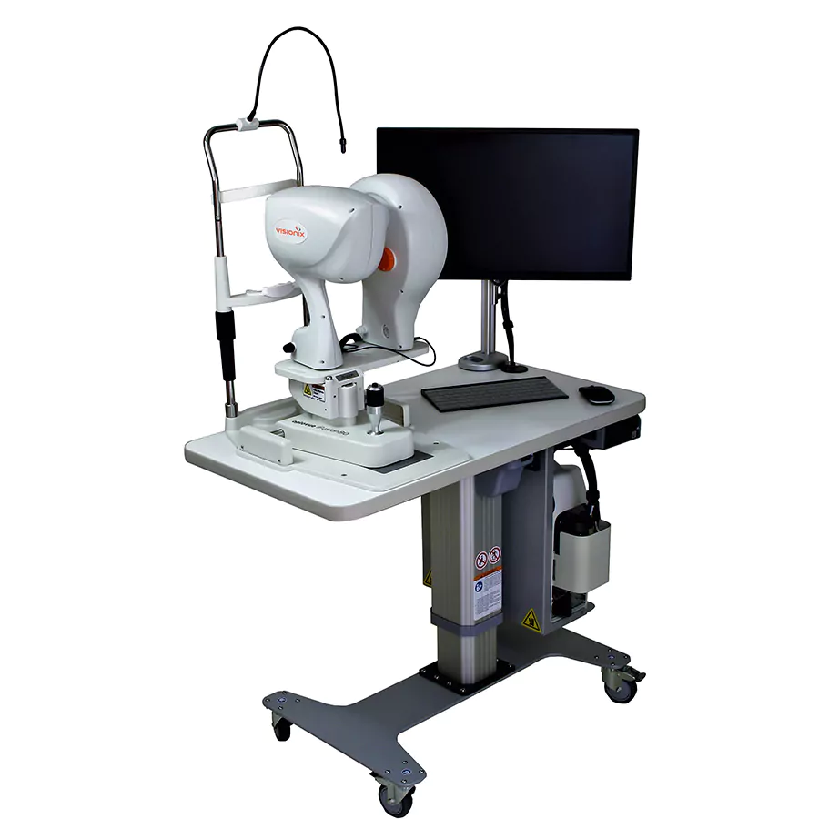
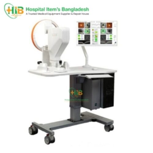
The complete retinal imaging iFusion Comprehensive OCT and Retinal Camera Platform
The iFusion OCT and retinal imaging system has all the same powerful clinical tools as the iVue with the added benefit of the iCam12 retinal camera. The iFusion transforms the way you assess the retina, optic disc and the cornea. Quantify the thickness of the retina, nerve fiber layer, ganglion cell complex (GCC) and the cornea. Track changes and predict trends in RNFL and GCC thickness and precisely measure angles to aid in disease diagnosis.

Pachymetry and Epithelial Thickness Mapping
Visualize and quantify 6mm of epithelial, stromal and total corneal thickness tonidentify areas of thickening or thinning related to dry eye disease, keratoconus, or previous refractive surgery. The Change Analysis report measures changes in thickness between visits.

Advanced GCC imaging
Advanced GCC imaging reveals ganglion cell and axon loss in optic nerve head disease. GCC thickness mapping improves clarity in structural change identification. Optovue’s exclusive FLV% and GLV% analyses increase GCC sensitivity and specificity.

iWellness Exam
The iWellness Exam is an Optovue exclusive that delivers a quick, easy OCT scan to promote better overall patient eye health. Its usefulness stems from a single, comprehensive report that depicts: retinal thickness and GCC thickness with normative comparison, symmetry analysis, FLV% and GLV%, proprietary Optovue GCC metrics that provide important information to aid in ocular disease diagnosis and management and eight high-resolution B-scans.

Retinal mapping and analysis
Retina mapping with normative comparison, retina change analysis and 3D retinal imaging with en face presentation are all depicted in high-resolution, easy-to-interpret colour reports.

Optic disc and RNFL assessment
Advanced capabilities include RNFL and GCC combination reports with normative comparison as well as RNFL and GCC trend analysis — both standard.
Comprehensive anterior segment analysis
Anterior segment capabilities include highly detailed reports for pachymetry mapping, anterior segment angle measurement.

3D and En Face imaging
3D and en face view provides multi-layer, high-resolution virtual dissection of the retina and optic disc, and depicts them in a way that preserves the retina’s natural curvature. This reduces distortion for simpler interpretation and enhanced 3D visual assessment.

iCam12 fundus camera
High-performance fundus camera that delivers posterior and anterior segment images with exceptional depth, 45° colour and red-free imaging, with image sharpening. The system software is very easy to learn and enables fast, flexible image review right out of the gate. With iFusion, you can quickly overlay iVue OCT images onto the fundus photos.

Easy to use software
The system software is very intuitive with helpful graphics and timely prompts that walk you through an exam. Most users are up to speed in a day.
Related products
-
Buy on WhatsApp
-
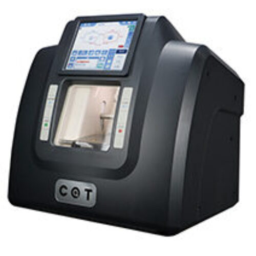
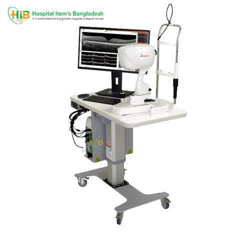
OCT-Optovue iVue 80
- Get Details
-
-
Buy on WhatsApp
-
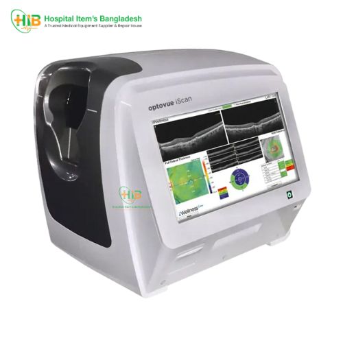
OCT-Optovue iScan 80
- Get Details
-
-
Buy on WhatsApp
-
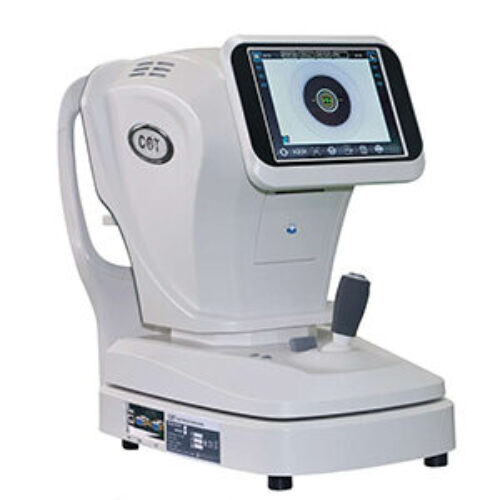
OCT with Angiogram
- Get Details
-
-
Buy on WhatsApp
-
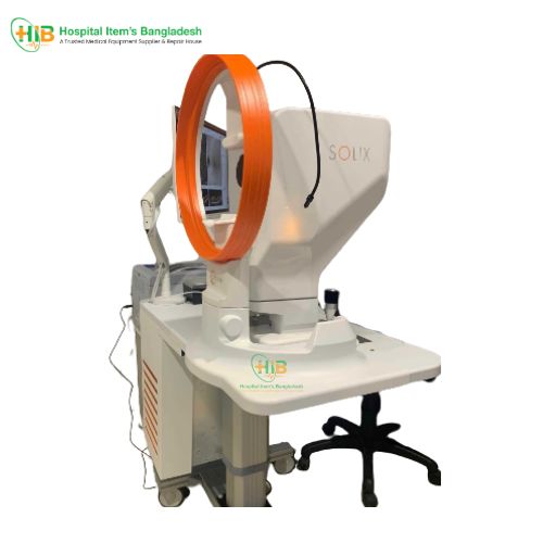
OCT-Optovue Solix SD-OCT
- Get Details
-


 Loading...

Reviews
There are no reviews yet.